Skip to content

Why Is a Bone Biopsy Procedure Needed?

Why Is a Bone Biopsy Procedure Needed? This question often arises after an X-ray, MRI, or CT scan shows an unusual area inside a bone. A scan can reveal a shadow, fracture, cyst, or mass. It cannot always identify the exact cause. A Bone Biopsy Procedure allows specialists to examine a small tissue sample under a microscope. The result may distinguish infection, benign growth, inflammatory disease, or cancer.

“Biopsy remains the most important step in establishing the diagnosis of a bone tumor,” explains Dr. Henry Jaffe, a pioneer in orthopedic pathology. His statement highlights an essential point: imaging guides suspicion, but tissue examination often provides confirmation. The procedure may use a needle under imaging guidance, or a small surgical incision. Doctors select the method according to the lesion’s location, size, and suspected behavior. Careful planning matters. A poorly placed biopsy can complicate later treatment.

The process is usually organized by an orthopedic oncologist, radiologist, and pathologist. Patients may receive local anesthesia, sedation, or general anesthesia. Some soreness or bruising can follow. Serious complications are uncommon, but bleeding, infection, and sampling errors remain possible. The sample can also be too small. That can be frustrating.

A Bone Biopsy Procedure is not automatically proof of cancer. It is a diagnostic tool, not a verdict. Doctors compare pathology findings with symptoms, blood tests, and imaging. Patients should ask who will perform the biopsy, how results will be reviewed, and when a second opinion is appropriate. Medical decisions need context. Even experienced teams sometimes reconsider the initial interpretation.
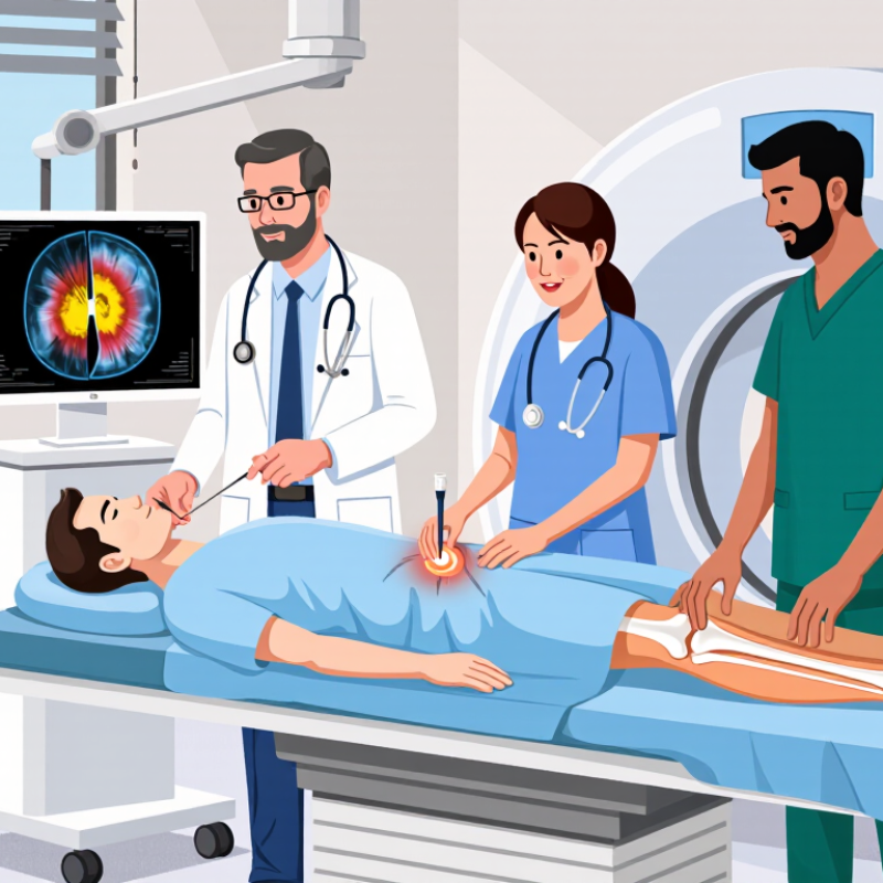
Why Is a Bone Biopsy Procedure Needed?

What Is a Bone Biopsy?

What Is a Bone Biopsy?

A bone biopsy is a procedure that removes a small bone or tissue sample for laboratory examination. A pathologist studies the sample under a microscope. The result can help distinguish infection, benign growths, inflammation, and cancer. Imaging scans may show an abnormal area, but they cannot always identify its exact cause.

Doctors usually perform a needle biopsy through the skin. Local anesthetic numbs the area, while computed tomography or ultrasound may guide the needle. Sometimes, a surgeon takes the sample through a small incision. The method depends on the lesion’s size, depth, location, and suspected diagnosis. It can feel like pressure, tapping, or brief soreness.

The American Cancer Society estimated about 4,040 new primary bone and joint cancer cases in the United States during 2025. These cancers remain uncommon, making accurate tissue diagnosis especially important. The American College of Radiology recommends image-guided biopsy when suitable, because planning can improve safety and diagnostic value.

A biopsy is not a perfect snapshot. The needle may miss the most informative tissue, and laboratory interpretation can require additional testing. Patients should ask how the biopsy site will be chosen and when results may arrive. Bleeding, infection, and temporary pain are possible, although serious complications are uncommon. A careful clinical history still matters.

Why Is a Bone Biopsy Procedure Needed?

A bone biopsy removes a small sample of bone or bone marrow so a laboratory can examine it under a microscope and perform other tests. It may help identify infection, cancer, unexplained bone lesions, or metabolic bone disease when imaging and blood tests cannot provide a definitive diagnosis.

Why Is a Bone Biopsy Recommended?

Why Is a Bone Biopsy Procedure Needed?

Why Is a Bone Biopsy Recommended?

A bone biopsy is recommended when imaging shows a suspicious or unclear bone lesion. X-rays, CT scans, and MRI can reveal size, location, and tissue changes. However, they cannot always identify the exact cause. The lesion may be benign, infectious, metastatic, or a primary bone tumor. A biopsy provides tissue for microscopic examination. This evidence often guides surgery, medication, or careful observation.

Primary bone cancer is uncommon. The National Cancer Institute’s SEER Cancer Stat Facts reports an estimated incidence of about 0.9 cases per 100,000 people annually in the United States. That rarity matters. Most suspicious bone findings are not primary bone cancer, but doctors still need a reliable diagnosis. A needle biopsy may collect a small core through the skin. Imaging guidance helps target the safest and most representative area.

The decision is not always tidy. A painful lesion may be harmless, while a painless lesion may still require testing. Biopsy can cause temporary soreness, bleeding, or infection, although serious complications are uncommon when specialists perform it. The American College of Radiology emphasizes planning the biopsy with the treating surgical team. Poor placement can affect later treatment. Patients should ask what the scan suggests, why tissue is needed, and how the result could change care. No test is perfect, and sometimes a repeat sample is necessary.

How Is a Bone Biopsy Performed?

Why Is a Bone Biopsy Procedure Needed?

A bone biopsy removes a small tissue sample for laboratory examination. Doctors may recommend it when scans show an unexplained lesion, infection, or possible bone cancer. The sample can clarify what imaging cannot. A trained radiologist or orthopedic surgeon performs the procedure, depending on the bone’s location and the suspected condition.

How Is a Bone Biopsy Performed?

Before the biopsy, the medical team reviews scans, medications, allergies, and bleeding risks. You may receive local anesthetic, sedation, or general anesthesia. The skin is cleaned carefully, then covered with sterile drapes. For a needle biopsy, the doctor guides a hollow needle through the skin and into the bone, often using CT or another imaging method. You may feel pressure, but sharp pain should be reported immediately.

The needle removes one or more small cores of tissue. An open biopsy uses a small surgical incision instead. It may be chosen when a larger sample is necessary. The wound is closed with stitches or a dressing, and pressure helps control bleeding. The specimen goes to a pathology laboratory for expert review.

The area may ache afterward.

Keep the dressing clean and follow the written instructions. Fever, increasing redness, drainage, heavy bleeding, or worsening pain requires prompt medical advice. A normal-looking wound does not always mean healing is complete. The exact procedure varies, and patients should ask about anesthesia, recovery time, and when results are expected.

What Conditions Can a Bone Biopsy Help Diagnose?

Why Is a Bone Biopsy Procedure Needed?

A bone biopsy removes a small tissue sample for laboratory examination. It is usually considered after X-rays, CT scans, MRI, or blood tests show an unexplained bone lesion. The procedure can help distinguish a benign growth from a primary bone cancer or cancer that has spread from another organ. It may also identify osteomyelitis, a deep bone infection, when cultures or imaging remain unclear. Sometimes, the answer is less dramatic. A lesion may reflect an inflammatory or metabolic disorder.

The U.S. National Cancer Institute’s SEER Cancer Stat Facts report shows that bone and joint cancers represent less than 1% of new cancer cases.

Their rarity makes accurate tissue diagnosis especially important. A pathologist can examine cell structure, while microbiology testing may detect bacteria or fungi in suspected infections. Biopsy results can also guide surgery, medication, or further imaging.

The sample is small, but its implications are not.

No biopsy is perfect. Sampling the wrong area can produce an incomplete answer, particularly in large or mixed lesions. Experienced radiologists and surgeons therefore plan the needle path carefully, avoiding areas that could complicate later treatment.

Patients should ask how the result will change management, because testing without a clear clinical question may add anxiety without adding useful information.

What Happens After the Biopsy?

After a bone biopsy, the care team usually checks the puncture site before discharge. You may have a small dressing, mild swelling, or tenderness around the area. Follow the written instructions carefully. Keep the wound clean and dry as advised. Avoid strenuous activity until your clinician gives permission.

Pain often improves within a few days. Prescribed or recommended medicine may help, but use it exactly as directed. Do not ignore increasing redness, warmth, drainage, fever, numbness, or severe pain. Contact your healthcare provider promptly if these signs appear. A small bruise can be normal. Heavy bleeding is not.

The tissue sample goes to a pathology laboratory for careful examination. Results may take several days or longer, depending on the tests required. Your doctor will arrange a follow-up appointment to explain the findings and discuss possible next steps. Sometimes the sample does not provide a clear answer, and another test may be considered. That possibility can feel frustrating. Recovery is not always perfectly predictable.

Write down new symptoms, medication questions, and concerns before your appointment. Bring someone you trust if the discussion feels overwhelming. Your experience matters, even when the clinical process seems routine.

Why Is a Bone Biopsy Procedure Needed? - What Happens After the Biopsy?

Data Dimension Clinical Topic Evidence-Based Information Typical Timing or Action
Purpose Why a bone biopsy is performed A bone biopsy removes a small sample of bone or bone marrow so it can be examined under a microscope. It may help identify the cause of unexplained bone pain, a suspicious imaging finding, infection, or an abnormal growth. Recommended when imaging tests and laboratory tests do not provide enough information.
Conditions Evaluated Possible diagnoses The procedure may help evaluate primary bone tumors, cancer that has spread to bone, bone infection, abnormal bone remodeling, and certain marrow-related disorders. Diagnosis depends on the biopsy result, imaging, medical history, and other laboratory findings.
Biopsy Type Needle biopsy A hollow needle is guided through the skin into the affected area to collect one or more tissue samples. Imaging guidance is often used to improve accuracy. Usually performed as an outpatient procedure when clinically appropriate.
Biopsy Type Open biopsy A surgeon makes a small incision to expose the bone and remove a tissue sample. This approach may be selected when a larger sample is needed or a needle biopsy is not suitable. May require regional or general anesthesia and can involve a longer recovery than a needle biopsy.
Before the Procedure Medication and health review The care team reviews allergies, medical conditions, bleeding risk, and current medicines. Blood-thinning medicines may require specific instructions from the treating clinician. Follow the individualized instructions provided before the appointment; do not stop prescribed medicine without medical advice.
During the Procedure Anesthesia and monitoring Local anesthetic is commonly used for needle biopsy. Sedation or general anesthesia may be used in selected cases. Vital signs are monitored during the procedure. Procedure length varies according to the biopsy site, technique, and number of samples required.
Immediately After Observation and discharge After the biopsy, the puncture or incision site is covered with a dressing. The patient is monitored until the effects of sedation or anesthesia have sufficiently worn off. Many patients go home the same day, although an overnight stay may be needed in some circumstances.
Expected Symptoms Pain, bruising, or tenderness Mild pain, tenderness, swelling, or bruising around the biopsy site can occur. These symptoms commonly improve gradually during the first few days. Use only the pain-relief medicines recommended by the care team and keep the site clean and protected.
Activity Returning to normal movement Activity restrictions depend on the biopsy location and technique. Strenuous exercise, heavy lifting, or putting weight on the affected limb may be limited temporarily. Resume normal activity gradually and follow the specific instructions provided at discharge.
Wound Care Protecting the biopsy site The dressing should be cared for according to the instructions provided. Keeping the area clean and dry can help reduce the risk of infection. Contact the care team if the dressing becomes soaked, the wound opens, or drainage continues.
Laboratory Analysis What happens to the sample A pathologist examines the tissue. Special stains, cultures, molecular tests, or other laboratory studies may be required depending on the suspected condition. Results are not always available immediately because additional testing may be necessary.
Results How findings are explained The clinician reviews the pathology report together with imaging studies, symptoms, and laboratory results. A biopsy can confirm a diagnosis, identify infection, or show that the sample is insufficient for a definitive conclusion. Follow-up timing varies according to the type of testing and the urgency of the clinical situation.
Possible Complications Risks to monitor Potential complications include bleeding, infection, persistent pain, nerve or blood-vessel injury, delayed wound healing, and, rarely, weakening or fracture of the affected bone. Risk depends on the biopsy site, technique, underlying condition, and overall health.
Warning Signs When to seek medical advice Urgent medical advice may be needed for heavy bleeding, increasing redness or warmth, pus-like drainage, fever, worsening pain, numbness, weakness, or difficulty moving the affected area. Seek emergency care for severe bleeding, breathing difficulty, fainting, or other rapidly worsening symptoms.
Follow-Up Care Next steps after results Further treatment may include antibiotics for infection, specialist consultation, surgery, cancer treatment, additional imaging, or observation, depending on the diagnosis. Attend the scheduled follow-up appointment even if the biopsy site feels normal.

Important: Recovery instructions and result timelines vary by procedure and individual health. The treating healthcare team’s instructions should take priority over general information.